Original Article – DOI: 10.33594/000000878
CPB (60): 386 - 407
Accepted: 27.05.2026 - Published: 31.07.2026
Macrophage Migration Inhibitory Factor (MIF) as a Central Regulator of Tumor Progression and Immune Evasion
bDepartment of Medical Biology and Genetics, Azerbaijan Medical University, Baku,
cDepartment of Oncology of Azerbaijan Medical University, Baku,
dDepartment of Pathology of Azerbaijan Medical University, Baku
Keywords
Abstract
Macrophage migration inhibitory factor has emerged as a central regulator of tumor progression and immune evasion. Initially identified as an inflammatory cytokine, it is now understood as a multifunctional signaling molecule that integrates hypoxia, metabolic stress, oncogenic activation, and tumor microenvironmental remodeling across diverse malignancies. Through pathways involving cluster of differentiation 74, cluster of differentiation 44, and C-X-C motif chemokine receptor 4, macrophage migration inhibitory factor promotes proliferation, survival, angiogenesis, epithelial-mesenchymal transition, invasion, and metastatic competence. It also shapes the tumor microenvironment by enhancing suppressive myeloid infiltration, macrophage polarization, cluster of differentiation 8-positive T-cell exhaustion, regulatory T-cell accumulation, and impaired antigen presentation. These combined effects position macrophage migration inhibitory factor as an immunometabolic mediator linking chronic inflammation with malignant adaptation and therapeutic resistance. Elevated signaling is associated with poor prognosis and reduced response to chemotherapy, radiotherapy, targeted therapy, and immune checkpoint blockade. Overall, macrophage migration inhibitory factor represents a mechanistically important and translationally relevant target whose clinical value will depend on precise biomarker-guided therapeutic integration.
Introduction
Macrophage migration inhibitory factor (MIF) is a key mediator at the intersection of inflammation and cancer biology. Tumor progression is increasingly recognized as being driven not only by intrinsic genetic alterations, but also by dynamic interactions between malignant cells and the stromal, vascular, metabolic, and immune components of the tumor microenvironment. In this sense, inflammatory cytokines have a major impact on the growth of tumors by influencing both antitumor immunity and the behavior of cancer cells. MIF is a cytokine, originally found to modulate innate immune responses, and has been increasingly acknowledged as a multifunctional molecule that is implicated in a variety of hallmarks of cancer, such as proliferation, survival, angiogenesis, invasion, and metastatic progression [1, 2]. Through its interactions with macrophages, stromal cells, vascular compartments, and immunological populations, the MIF cytokine family has been demonstrated to promote microenvironmental remodeling in addition to its direct action on tumor cells. These observations support the view that MIF functions not merely as a marker of inflammatory activation, but also as an active mediator of tumor progression [3].. There has also been a growing interest in the role of MIF in tumor immunity. Multiple studies have indicated that MIF can participate in the immunosuppressive mechanisms in the tumor microenvironment through mechanisms such as antigen presentation, recruitment of suppressive myeloid cells, dysfunction of T cells, and expansion of regulatory immune cells. In cancer, MIF may promote a switch from chronic inflammatory signaling to chronic immune suppression by these mechanisms. The relevance of MIF in translation has further grown by studying its receptor network and related family members. CD74 has become an important signaling partner with prognostic and immunological implications in various malignancies, and CD44 has been identified as a signaling component of the MIF-CD74 receptor complex [4, 5]. Furthermore, D-dopachrome tautomerizes (DDT, also known as MIF-2) and has expanded the functional complexity of the MIF signaling family [6, 7]. The evidence in this field so far indicates that the MIF axis should not be viewed only as a single cytokine axis, but rather as an integrated signaling network, especially in the context of biomarker development and therapeutic targeting [8, 9]. Evidence also suggests that MIF signaling is associated with aggressive tumor biology in several malignancies, including melanoma, breast cancer, lung cancer, and broader pan-cancer contexts. High MIF activity has been linked to immune changes, macrophage-associated phenotypes, and poor clinical features in several tumor systems [10, 11]. In the same way, in melanoma and thoracic malignancies, MIF is a signaling molecule that may play a role in tumor progression and drug resistance [12, 13]. Furthermore, pan-cancer analyses indicate that MIF could serve as a more general oncologic biomarker with immune associations [14]. In this context, this review is aimed at exploring the function of MIF in the regulation of tumor progression and immune evasion. Special focus is devoted to the molecular organization of the MIF signaling network, its involvement in the intrinsic and microenvironment-dependent processes in tumor cells, and its newfound role in immunometabolism adaptation and therapeutic resistance [15]. Furthermore, the translational implications of MIF for biomarker development, therapy responsiveness, and future precision-oncology strategies are addressed.
Objectives of the study:
- 1. To examine the molecular biology and signalling architecture of MIF and the MIF family in cancer, with particular emphasis on unconventional secretion, CD74-centred receptor complexity, CXCR4 interaction, and the overlapping but distinct roles of D-DT/MIF-2.
- 2. To evaluate the role of MIF in tumor progression and immune evasion across cancer systems, focusing on tumor-cell proliferation, survival, epithelial-mesenchymal transition, metastasis, tumor microenvironment remodelling, suppressive myeloid recruitment, T-cell dysfunction, and immunometabolism adaptation. 3. To assess the translational significance of the MIF axis in oncology, including its value as a biomarker and its therapeutic potential in relation to therapy resistance, immunotherapy responsiveness, and biomarker-guided precision treatment strategies.
Molecular Biology of MIF and the MIF Family
Discovery, structural organization, and unconventional secretion
MIF is a highly conserved multifunctional protein that plays important roles in inflammation, immune regulation, cellular stress responses, and cancer biology. MIF is a cytokine classified as inflammatory but is now known to be a member of a larger signaling family that includes D-dopachrome tautomerase (D-DT/MIF-2) and has both common and distinct biological functions [16]. One distinctive feature of MIF biology is its unconventional secretion pathway. In contrast to most classical cytokines, MIF is not secreted via the endoplasmic reticulum-Golgi secretory pathway. Rather, the intracellular MIF can be mobilized and quickly released upon cellular stress conditions. This mechanism can be important in situations of sustained extracellular release of stress-associated mediators, such as in tumors where oxygen levels, oxidants, metabolic instability, and chronic inflammatory signaling are all present.
Receptors and co-receptors: CD74-centered signaling complexity
The biological effects of MIF are highly dependent on receptor context, in different tissues and disease conditions. In addition to its classical role of antigen presentation, CD74 has been identified as a major receptor for MIF and has become an important signaling molecule in the biology of cancer. CD74-dependent MIF signaling has been associated with cell survival, immune regulation, and tumor progression in tumor systems [17]. Further complicating MIF signaling are interactions with other co-receptors and chemokine-associated receptor systems. MIF has been demonstrated to interact functionally with CXCR4, which has led to the idea that MIF signaling shares characteristics with chemokine-mediated signaling pathways [18]. Moreover, CD74 was found to be a functional receptor for MIF on activated CD4+ T cells, further expanding the role of MIF signaling in adaptive immune regulation and tumor-associated immune remodeling [19]. These findings suggest that MIF signaling is regulated by multiple receptors, which can have a variety of effects on inflammation, oncogenesis, and immune regulation.
D-DT/MIF-2 overlaps with MIF and conformational regulation
The closest homolog of MIF, D-dopachrome tautomerize (D-DT, also known as MIF-2), is an important member of the MIF signaling family. While there are some structural and functional similarities between D-DT and MIF, the present evidence does not suggest that D-DT is merely functionally redundant. Rather, there seems to be overlap and divergence between MIF and D-DT signaling, adding more complexity to the biological and therapeutic interpretation of this pathway. Conformational regulation also seems to be a key aspect of D-DT biology, and pathway activity might be regulated not just by alteration of expression levels, but also by dynamic structural variation of the protein [20]. Oxidative stress, receptor availability, and microenvironmental conditions may be particularly important in cancer settings, which could impact downstream signaling behavior. Thus, DDT can be viewed as a distinct mechanistic member of the MIF signaling family in terms of considering pathway biology and therapeutic targeting approaches [21]. Table 1 provides an overview of the primary ligands, receptors, signaling partners, and downstream signaling pathways of MIF-family signaling in cancer, highlighting the complexity of the receptor assemblies for CD74-mediated signaling, the signaling interaction of D-DT/MIF-2, and the roles of the signaling pathways in tumor progression, immune remodeling, and therapeutic targeting.
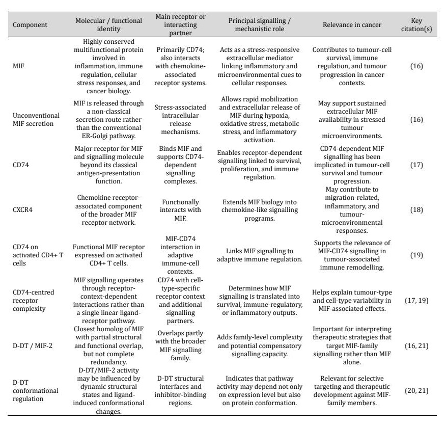
Table 1: Core molecular architecture of the MIF signaling system in Cancer
Upstream Regulation of MIF in the Tumor Context
Hypoxia, metabolic stress, and oncogenic control of MIF
Rather than representing a constitutive activation state, microenvironmental stress conditions seem to play a significant role in the expression of MIF in tumors. Aggressive tumors are defined by hypoxia, nutrient limitation, oxidative stress, and chronic inflammatory signaling, which can lead to upregulation of MIF as one of a set of mechanisms for cellular stress-adaptation. The MIF promoter polymorphism (rs755622) has been linked to immune activation in glioblastoma, indicating that genetic variation of the MIF regulation could affect the interaction with the tumor microenvironment [22]. This regulatory variation could influence the response of tumors to hypoxia- and inflammation-related transcription. Further experimental data from oral squamous cell carcinoma (OSCC) suggest that MIF is involved in stress-adaptation pathways. In this case, UVB-induced apoptosis and reactive oxygen species buildup were shown to be enhanced by MIF knockdown, indicating that MIF-mediated signaling may be involved in cellular defense under oxidative stress (23). Taken together, these results indicate that the expression of MIF seems to be tightly coupled to stress responses associated with tumors and helps to adapt malignant cells to metabolic and oxidative stress.
Inflammatory signalling, infection-associated stress, and tumor-promoting induction
MIF is also controlled by inflammatory and infection-related signaling pathways, which are often associated with tumor progression. MIF induction has been associated with chronic exposure to cytokines, activation of NF-kB, alterations in glucocorticoid signaling, and persistent cellular stress, which all involve long-term inflammatory adaptation within tumors. This correlation is seen especially in virus-related cancers. A role for MIF in HPV-driven head and neck squamous cell carcinoma in the remodeling of the cytokine microenvironment has been proposed, as HPV infection has been linked to increased MIF release (24). In a larger context, these findings suggest that in some tumor circumstances, MIF signaling may play a role in the interaction of infection, inflammation, and carcinogenic transformation (25). Collectively, available evidence suggests that MIF is a downstream mediator of tumor progression as well as a stress-responsive mediator that is regulated by microenvironmental conditions of inflammation and infection.
Epigenetic and non-coding RNA regulation of MIF expression
Apart from environmental and inflammatory regulation, the expression of MIF also seems to be regulated by epigenetic mechanisms and non-coding RNA-associated regulatory networks. These higher-order regulation processes may have special relevance in cancer due to their ability to fix adaptive phenotypes under chronic microenvironmental stress and therapeutic pressure. Epigenetic processes such as chromatin-associated and transcription regulatory mechanisms are possible ways by which the activity of MIF signaling may be affected in cancer and inflammatory diseases, as previously suggested (26). At the same time, MIF-associated signaling pathways may be post-transcriptionally regulated by non-coding RNA pathways. The significance of post-transcriptional control in MIF-associated cancer biology is demonstrated by the fact that microRNA-mediated regulation has been shown to be involved in the regulation of MIF synthesis and proliferation in gastrointestinal cancer cells (27). These findings collectively imply that environmental stress, inflammatory signaling, epigenetic remodeling, and post-transcriptional regulation are all part of a complex and dynamic regulatory network that controls MIF expression.
Core Signalling Networks Downstream of MIF
CD74-dependent signalling and receptor complex formation
A central feature of MIF signaling in cancer is the context in which the receptor is found, especially MIF associated with CD74. CD74 is not only a binding receptor but also an active signaling platform that can be involved in microenvironment-associated cellular responses, proliferation, and survival. The connections between cell-surface CD74 and MIF in melanoma, which have been shown to support the tumor cells' survival, thereby illustrate how receptor-mediated interactions contribute to cancer-cell persistence (28). Receptor-associated signaling patterns are also described in lung cancer, where spatial downregulation of CD74-related signatures has been linked to the development of invasive features in part-solid lung adenocarcinoma (29). Altogether, these data indicate that CD74 expression and spatially associated CD74 signatures might influence tumor behavior and serve as a biological hallmark of receptor-associated signaling in tumor systems.
MAPK/ERK and PI3K/AKT/mTOR pathway activation
The most well-described downstream signaling pathways stimulated by MIF include the MAPK/ERK and PI3K/AKT pathways, two major pathways involved in the regulation of the proliferation, survival, and metabolic adaptation of tumor cells. The activation of the PI3K/Akt pathway by MIF signaling has been associated with gastric cancer, in which MIF has been shown to regulate the proliferation of the tumor cells (30). In the same way, MIF signaling in CRC has been linked to increased malignant progression and activity of oncogenic signaling pathways (31). The activation of these signaling cascades can affect several other downstream processes, such as resistance to apoptosis, cell cycle progression, and adaptation to stress. MIF may therefore function as an upstream coordinator that connects external inflammatory signals with intracellular survival and growth programs due to the substantial interaction between the PI3K/AKT/mTOR and MAPK/ERK pathways with other oncogenic signaling pathways.
Integrative signaling crosstalk, redox adaptation, and immunometabolic rewiring
MIF signaling beyond the receptor seems to be involved in a complex network of inflammatory, angiogenic, and immunometabolism regulatory pathways, instead of a linear signaling cascade. In colorectal cancer, Hsp90-mediated stabilization of MIF has been linked to continuous tumor progression, recruitment of macrophages, and angiogenesis, indicating that the interplay between MIF signaling and chaperone biology is linked to stromal communication (32). Further research in lung cancer has revealed that receptor molecular contexts, such as the CD74-ROS1 fusion transcript in non-small cell lung carcinoma, may be associated with biologically distinct signaling contexts (33). All this evidence is in favor of MIF signaling being involved in several regulatory pathways, such as inflammatory transcription factors NF-κB and AP-1, survival pathways such as STAT3, and redox-sensitive adaptive programs. Thus, MIF signaling is now regarded as part of a concerted signaling network involved in the inflammatory adaptation and microenvironmental remodeling in tumors. The key players in the MIF signaling axis in cancer are summarized in Fig. 1. This figure depicts the major ligands MIF and D-DT/MIF-2, the main receptor-associated components CD74 and CXCR4, and the activation of signaling and redox-associated stress pathways. These signaling have been associated with tumor proliferation, survival, EMT/metastasis, angiogenesis, macrophage recruitment/polarization, immune suppression, T-cell dysfunction, therapy resistance, and immunometabolism adaptation.
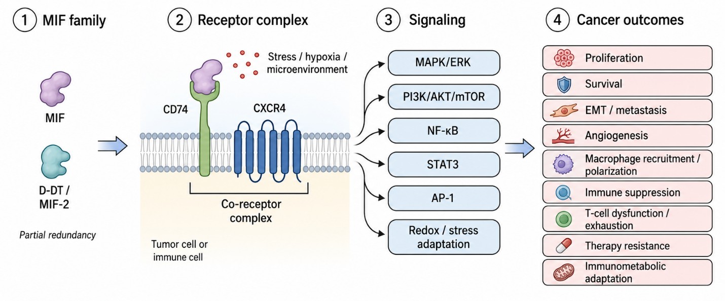
Fig. 1: The MIF signaling axis in cancer: ligands, receptor complexes, and downstream pathways-
MIF as a Driver of Tumor Cell-Intrinsic Malignancy
Proliferation, cell-cycle progression, and survival advantage
Through the stimulation of growth-associated signaling pathways, it is becoming evident that MIF directly contributes to the growth and survival of tumor cells. The idea that MIF is actively involved in the regulation of tumor cell growth rather than only acting as a secondary inflammatory marker is supported by the fact that inhibition of MIF in bladder cancer resulted in a decrease in cell growth and cytokine expression (34). The same has been observed in pancreatic cancer, where the overexpression of MIF in tumor tissues has been correlated to tumor progression and malignant behavior (35). These observations collectively indicate that MIF-related signaling may help to maintain the viability of tumor cells and promote cell-cycle progression in the hostile microenvironment.
EMT, invasion, metastatic competence, and malignant plasticity
MIF signaling has also been linked to epithelial-mesenchymal transition (EMT), invasion, and metastatic progression. MIF has been demonstrated to induce changes related to EMT and enhance tumor aggressiveness in pancreatic ductal adenocarcinoma, and has been associated with the development of invasive and metastatic cellular traits (36). These findings indicate that MIF could be involved in wider malignant reprogramming processes, given its link to EMT, as well as the migratory capacity and therapy adaptation of cells. Finally, CD74-MIF signaling in melanoma has been linked to tumor growth and patient survival, which reinforces the role CD74-MIF signaling can play in determining the behavior of tumor cells under different biological conditions (37). Together, these studies suggest that MIF-mediated signaling can contribute to the shift to more aggressive and therapy-resistant malignant conditions.
Stress adaptation, tumor initiation pressure, and therapy-linked malignant persistence
MIF could also play a role in the persistence of the tumor by contributing to adaptation to environmental and genotoxic stresses. The MIF signaling family may be involved in the adaptation of malignant cells to DNA-damaging conditions in tumor initiation and progression, as both MIF and D-DT are implicated in UVB-induced carcinogenesis in non-melanoma skin cancer (38). Similar findings have been reported in lung cancer, where the development of several primary lung adenocarcinomas has been linked to MIF-associated signaling, indicating a possible function for MIF in tumor-promoting field effects and long-term microenvironmental adaptability (39). Together, this shows that MIF signalling contributes to cellular stress tolerance, persistence under adverse microenvironmental conditions, and maintenance of malignant phenotypes during disease progression.
MIF in the Tumor Microenvironment
MIF-mediated remodelling of myeloid infiltration in the tumor microenvironment
MIF also plays a central role in the organization of the tumor microenvironment by influencing the recruitment and localization of myeloid-cell populations that are associated with tumor progression. In addition to its role as a soluble inflammatory mediator, MIF seems to be involved in creating an immunosuppressive microenvironment around malignant cells. In hepatocellular carcinoma, MIF was found to contribute to tuning the composition of phagocyte-lineage cells within the tumor by regulating mononuclear phagocyte infiltration (40). This finding has important implications for tumor-associated immune remodeling because the tumor microenvironment's functional characteristics depend on the ratio of pro-inflammatory to tumor-promoting myeloid cells. Furthermore, it has been proposed that tumor-associated macrophages play a critical role in the tumor microenvironment in HCC and may encourage the advancement of the disease and resistance to treatment (41).
MIF-CD74 signaling, macrophage polarization, and immunosuppressive remodelling
Recent findings also indicate that MIF is involved in receptor-mediated signaling pathways that are responsible for the polarization of macrophages and the overall immunosuppressive remodeling. Single-cell RNA-sequencing research suggests that the MIF-CD74 axis may play a role in the immunosuppressive reprogramming of TAMs in the tumor microenvironment in gastric cancer (42). As changes in macrophage phenotypes can greatly impact the behavior of a tumor, disruption of signaling pathways associated with MIF may alter the balance of inflammatory and tumor-promoting immune contexts. Sphingosine-1-phosphate (S1P) further highlights the importance of MIF signaling in immune modulation and vascular remodeling in the setting of colorectal cancer. A molecule involved in MIF signaling has been linked to the stimulation of angiogenesis and macrophage polarization (43). This indicates that MIF is part of a complex signaling network that includes inflammatory, stromal, and receptor-dependent pathways, which are all important in tumor adaptation and progression.
MIF-centered immunoregulatory networks and suppressive cell recruitment
Beyond macrophage remodeling, MIF also seems to be involved in a large network of immunoregulatory events of the tumor microenvironment, such as the recruitment and maintenance of suppressor immune-cell populations. MIF has been demonstrated to induce the growth and metastasis of tumors by inducing myeloid-derived suppressor cells (MDSCs), implying that MIF plays a role in the induction of local immune suppression (44). MDSCs are known as significant mediators of defective anti-tumor immunity, and these data suggest that they could be relevant to tumor progression and therapeutic resistance. Similarly, MIF is secreted by cancer stem cells and has a stimulatory effect on MDSC function and immune evasion in glioblastoma (45). Thus, further supporting a role for MIF in suppressive immune-cell programming in the tumor microenvironment. In sum, current evidence suggests a role of MIF in the localization and functional programming of immune-cell subsets involved in tumor-associated immune remodeling and immune evasion. Fig. 2 summarizes MIF's function in immune evasion and tumor microenvironment remodeling. Hypoxia, inflammation, oxidative stress, and metabolic stress can all induce MIF secretion by tumour cells, and subsequent downstream signaling through the MIF-CD74 axis and other immunomodulatory pathways is shown.
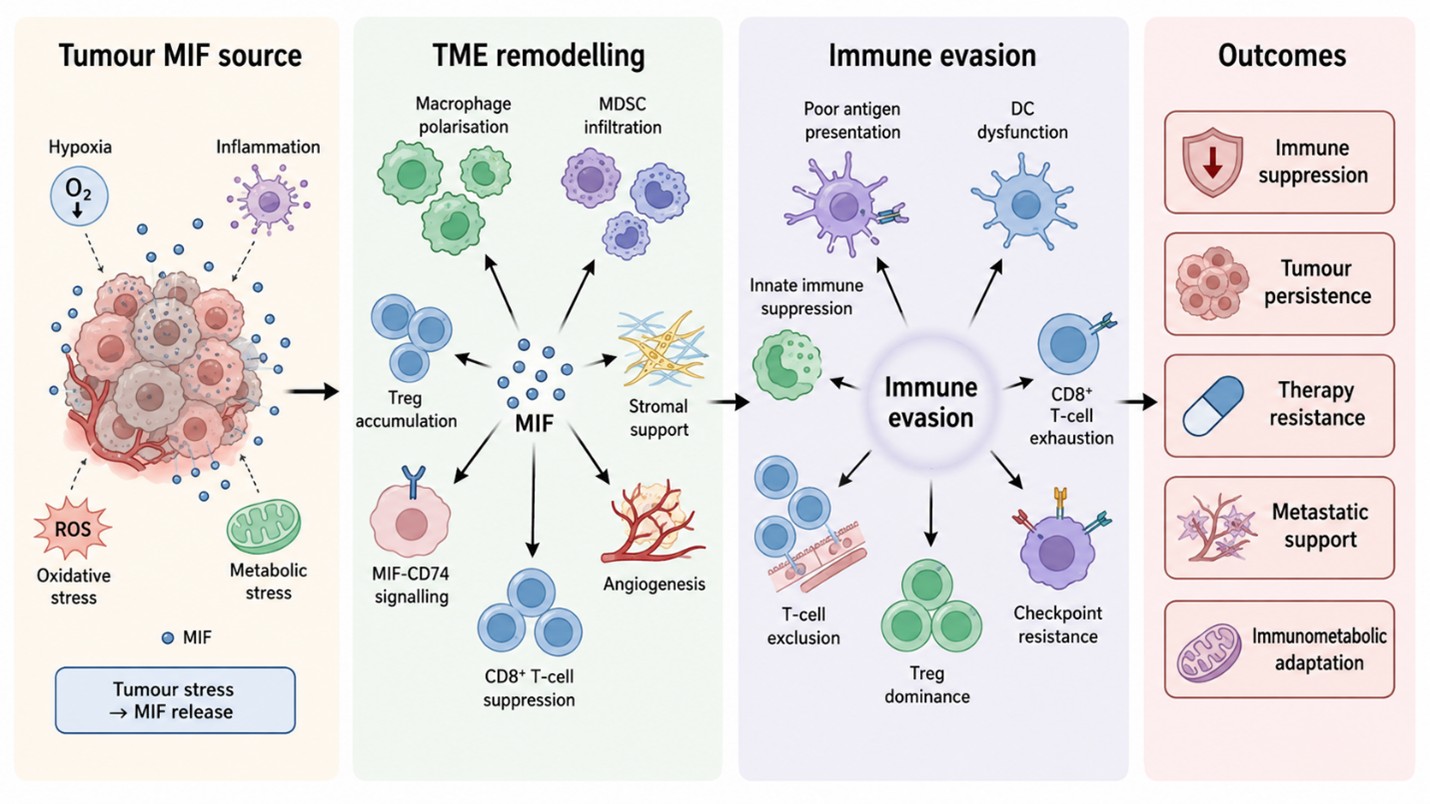
Fig. 2: MIF-driven remodelling of the tumor microenvironment and immune evasion.
MIF as a Central Regulator of Immune Evasion
Suppression of innate antitumor immunity and antigen presentation failure
Accumulating evidence suggests that MIF plays a role in the early events of tumor-immune interaction in immune evasion, by affecting innate antitumor immunity and antigen-presentation mechanisms. The regulation of immune cell recruitment and functional programming in the tumor microenvironment is one proposed mechanism. In head and neck squamous cell carcinoma, MIF is linked to a decrease in the intratumoral accumulation of anti-tumoral immune-cell populations and to the creation of an intratumoral immunosuppressive environment, indicating that MIF signaling might be involved in immune remodeling that is supportive to the tumor (46). Further, CD74-related immune signals can also impact therapeutic sensitivity, as well as tumor immune recognition. In the context of cervical cancer, targeting CD74 from TAMs enhanced the therapeutic effect of neoadjuvant chemotherapy in combination with PD-1 inhibition, underscoring the importance of CD74-mediated immune regulation in therapeutic responses against cancer (47). Together, these observations argue that MIF/CD74-mediated signaling events can help to subvert immune surveillance and immune-recognition events in the microenvironment of tumors.
T-cell exhaustion, exclusion, and checkpoint-linked immune dysfunction
MIF has also been related to several different types of T-cell dysfunction associated with tumor immune evasion. There is some evidence that high activity of MIF leads to T-cell exclusion, poor effector function, and suppressive immune conditions within tumor tissues. MIF is a clinically relevant and functional in vitro biomarker in multiple myeloma (MM), where it is associated with multiple disease-associated immune and survival-related states (48). Similarly, research on targeting MIF in head and neck squamous cell carcinoma (HNSCC) has shown that MIF may play a role in reducing T-cell immunosuppression in these tumors, suggesting that the MIF pathway is involved in promoting suppressive immune environments in tumors (49). Moreover, the simultaneous inhibition of MIF and PD-1 signaling has been shown to improve the response to antitumor immune responses, suggesting possible functional similarities between MIF-associated signaling and immune resistance mechanisms via checkpoint inhibitors (50).
Regulatory T cells and immunosuppressive cytokine landscapes
MIF not only affects myeloid populations and effector T cells, but it also seems to affect the biology of regulatory T cells and the greater immunosuppressive cytokine networks in tumors. This facet of MIF signaling is of particular interest because it connects chronic inflammatory signaling with the maintenance of a suppressive immune state, but not of productive anti-tumoral immune responses. According to experimental research, regulatory T cell enrichment and function in the tumor microenvironment are influenced by MIF-associated receptor signaling (51). All of these findings suggest that MIF's coordinated effects on immune-suppressive cells, inflammatory pathways, and cytokine-mediated immune control may contribute to the development of an immunologically tolerant tumor microenvironment (TME).
MIF, Tumor Metabolism, and Immunometabolic Reprogramming
Glycolysis, lactate-rich microenvironments, and metabolic competition
There are many similarities between tumor metabolism and immune suppression, and increasing data suggest that MIF might be involved in this relationship via its connection with the glycolytic and immunosuppressive tumor microenvironments. Single-cell transcriptional analysis of pancreatic ductal adenocarcinoma has revealed that immunosuppressive microenvironmental states emerge during the evolution of metastatic progression, suggesting that metabolic and immune remodeling progress in parallel during tumor evolution (52). The glycolytic metabolism causes lactate accumulation and extracellular acidification, which have the ability to activate anti-tumor immune responses and induce adaptive survival mechanisms within the tumor cells. The metabolic regulation associated with MIF has also been explored in pancreatic cancer, revealing that MIF interaction with NR3C2 is linked to MAPK–ERK and AP-1 pathways in the regulation of reprogramming of glucose metabolism (53). Taken together, these observations indicate that MIF signaling should be viewed in the context of immunometabolism in a broader sense, where competition for nutrients, metabolic changes, and Tumor cell fitness and immune cell function are both impacted by microenvironmental stress.
Mitochondrial stress, lipid imbalance, and myeloid-cell dysfunction
MIF-induced immunometabolism remodeling might not be limited to glucose metabolism but also involve mitochondrial stress responses, lipid metabolism, and functional regulation of myeloid-cell populations. Single-cell transcriptomic analysis in pancreatic cancer has revealed types of tumor-associated macrophages with prognostic significance, emphasizing the role of restructuring the myeloid cells in the progression of the disease (54). These changes can correlate with changes in fatty-acid metabolism, mitochondrial adaptation, and oxidative-stress response, which can all result in suppressive immune phenotypes. Further, obesity can induce changes in the metabolism of the tumor microenvironment and promote the inhibition of antitumor immunity, thus further supporting the overall idea of how host metabolic state might affect tumor-associated immune remodeling (55). These observations indicate that the MIF signaling network may be integrated into a broader metabolic network that includes oxidative stress, lipid-rich microdomains, mitochondrial adaptation, and all that influences tumor-promoting immune conditions.
MIF as an immunometabolism checkpoint candidate
MIF signaling is involved in both metabolic adaptation and immune regulation, and it has been suggested as an immunometabolism checkpoint-associated pathway in cancer. This is especially true in pancreatic cancer, where transcriptional and epigenetic programs are tightly associated with immune-microenvironment organization. In pancreatic ductal adenocarcinoma, for example, the expression of m6A regulators has been linked to aspects of the immune microenvironment and prognosis, suggesting that post-transcriptional regulatory mechanisms may help stabilize metabolically adapted and immune-evasive states of cells (56). The idea that MIF-related signals should be comprehended in an integrated metabolic and immune-regulatory context is supported by the fact that immunometabolism interactions within the tumor microenvironment can influence the viability of tumor cells, the activity of immune cells, and their response to therapy (57). Additional mechanistic and clinical investigations will be needed, however, to establish the extent to which MIF can be considered to be a clinically applicable immunometabolism regulator in cancer.
MIF Across Major Solid and Hematologic Malignancies
Glioblastoma, CNS tumors, and thoracic malignancies
The increasing amount of data suggests that MIF has a role in the biology of glioblastoma (GB) and other cancers of the central nervous system through its activities in tumor maintenance, tumor microenvironment adaptability, and treatment resistance. As a result, MIF's potential as a therapeutic target in neuro-oncology is gaining attention, particularly in relation to experimental and translational research (58). MIF-associated signaling has also been shown to be involved in tumor progression in thoracic malignancies; however, there are spatially and biologically heterogeneous mechanisms. Pharmacological inhibition of MIF has been demonstrated to inhibit the growth and mobility of lung cancer cells, indicating that MIF is involved in the malignant progression and adaptive behavior of tumor cells (59). Together, these results suggest that pathways involved in MIF may be of prognostic and therapeutic value in CNS and thoracic malignancies with significant immune and microenvironmental characteristics.
Colorectal, gastric, hepatobiliary, and pancreatic cancers
High levels of MIF signaling have been associated with adverse clinicopathologic parameters in gastrointestinal and hepatobiliary tumors, but the major mechanisms seem to vary with the tumor type. In gastric cancer, the higher levels of MIF were related to increased angiogenesis and stage of the disease, suggesting that MIF activity relates to the progression of the disease (60). Single-cell and transcriptomic studies have also shown that the progression of gastric cancer and liver metastasis is driven by complex interactions between epithelial, stromal, and immune-cell populations, in which MIF-related signaling might be involved in microenvironmental remodeling (61, 62). The clinical significance of myeloid-cell regulation becomes more evident in the case of pancreatic cancer, where the presence of M2-polarized tumor-associated macrophages (TAMs) is correlated with aggressive tumor behavior, indicating that macrophage-related immune phenotypes are important in the pancreatic tumor microenvironment (TME) (63). This is particularly relevant in pancreatic ductal adenocarcinoma, where disease behavior and therapeutic responses are heavily influenced by stromal remodeling, metabolic stress, and immune suppression.
Melanoma and genitourinary malignancies
MIF signaling also seems to be a significant factor in melanoma biology in terms of tumor-cell signaling and microenvironmental regulation. MIF-CD74 interaction has been demonstrated to control programmed death ligand 1 (PD-L1) expression in melanoma cells, indicating that receptor-associated MIF signaling may play a role in immune regulation and mechanisms of tumor immune escape (64). These results reinforce the idea that MIF-related pathways might play multiple roles in both immune escape and facilitation of tumor-promoting microenvironments in melanoma. In other types of cancer, MIF-mediated signaling has also been studied in the context of tumor progression and therapeutic targeting. Specifically, oxidized MIF has been suggested as a druggable disease-associated form of MIF in cancer, thus bolstering the overall translational relevance of MIF-directed approaches (65). Despite the differences in their biological backgrounds, all of these studies point to the possible adaptability of MIF as a potential therapeutic target and biomarker candidate in many cancers.
Hematologic malignancies and cross-cancer synthesis
In haematologic malignancies, MIF seems to contribute to disease persistence, not only by direct effects on malignant-cell growth, but also by modulating immune and stromal microenvironments. Despite the fact that there are fewer studies in the field of hematologic tumors than in some solid tumors, the data indicate that MIF-related pathways might play roles in disease-associated survival, immune dysfunction, and adaptation of the microenvironment. Furthermore, oxMIF-targeting radioimmunotherapy has been shown to induce considerable tumor regression in animal cancer models, underscoring the therapeutic potential of MIF-targeting approaches in specific cancer types (66). The main functional activities of MIF in tumor progression and immune evasion are listed in Table 2. The table summarizes the roles of MIF in the tumor intrinsic mechanisms of proliferation, survival, EMT, metastasis, and angiogenesis, and in the remodeling of the tumor micro-environment by MIF-induced macrophage polarization, recruitment of MDSC, stromal support, and accumulation of Tregs.
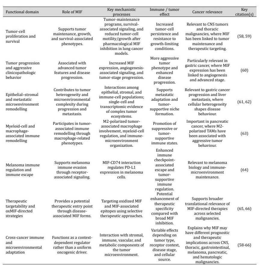
Table 2: Functional roles of MIF across tumor progression and immune evasion
MIF and Resistance to Anticancer Therapy
Chemoresistance and resistance to targeted therapy
The growing evidence indicates that MIF is involved in therapeutic resistance by its role in survival signaling pathways and adaptive receptor-associated networks that maintain the survival of tumor cells during therapy. The role of MIF has been explored as a therapeutic target in colorectal cancer, indicating that MIF-related signaling pathways may be involved in the persistence of the tumor and adaptive escape to therapy (67). Other gastrointestinal malignancies have shown MIF-associated pathways to be significant in treatment responsiveness. In pancreatic cancer, targeting mast-cell activation has been found to improve chemotherapy response (68). This suggests a possible correlation between MIF signaling, adaptation of the tumor microenvironment, and therapeutic resistance. These results suggest that MIF could play a role in resistance to conventional anti-cancer strategies.
Radioresistance, stemness, and adaptive survival
MIF signaling has also been linked to resistance to radiotherapy (RT) via cellular plasticity and survival-associated phenotypes. The MIF inhibitor 4-IPP suppressed stemness-related phenotype and mesenchymal trans differentiation after irradiation in glioblastoma multiforme, indicating that the MIF-associated pathways might be involved in adaptive response to irradiation-induced stress (69). The observed associations with radio resistance may extend to MIF's capacity to maintain therapy-tolerant cellular states, as these stem-like tumor-cell populations can repopulate tumors after therapy. It is therefore possible that under certain circumstances, MIF-dependent signaling could be involved in minimal residual disease and tumor persistence after treatment.
Resistance to immune checkpoint blockade and hyper-progressive disease
The role of MIF in therapy resistance is receiving increasing attention because of its association with tumor-associated immune dysfunction and treatment-adaptive microenvironmental remodeling. These findings further support the therapeutic relevance of MIF-directed immune modulation (70). Overall, these findings indicate that targeting MIF could be achieved not only by blocking the signaling activity of MIF but also by redirecting immune responses against MIF-associated aspects of tumor-supportive mechanisms. This interpretation is significant for helping understand how MIF can promote immune suppression, stromal remodeling, and adaptive survival, which can lead to decreased sensitivity to therapy. While these results are still in the preclinical stage, they provide a basis for further exploring MIF and related pathways as potential regulators of therapeutic responsiveness and immune-treatment resistance.
CD74-linked response prediction and relapse-associated therapeutic failure
Since MIF signaling is heavily reliant on interactions with the receptor, therapeutic responsiveness may be dependent on the abundance of ligand and also the composition and pathway organization of the receptor. By causing microglial M1 polarization, blocking the MIF-CD74 axis has been demonstrated to increase the efficacy of radiotherapy in non-small cell lung cancer brain metastasis. This suggests that receptor-mediated MIF signaling may be targeted in particular tumor types to decrease metastasis and improve treatment outcomes (71). The findings suggest that malignancies with particular immune-microenvironment and therapeutic-response characteristics may be identified by focusing on the MIF-CD74 axis. Taken in the context of the association of MIF signaling to chemoresistance, radio resistance, and checkpoint-associated immune dysfunction, these observations indicate that a wider role for MIF-associated pathways in tumor persistence and treatment adaptation must be considered. Hence, the MIF-CD74 signaling axis could be a resistance mechanism as well as a possible biomarker for therapy-response stratification for certain malignancies.
Therapeutic Targeting of MIF
Direct inhibition of MIF and the problem of target complexity
The role of MIF in the survival, immune modulation, and microenvironmental adaptation of tumor cells has led to a growing interest in therapeutic targeting of this molecule. Small molecule inhibitors and isoform-selective approaches are of particular interest, but the structural and functional complexity of the MIF signaling network brings important challenges to therapeutic development. Experimental studies indicate that MIF can be a potentially relevant therapeutic target in glioblastoma by applying inhibitor-based strategies (72). Another translational interest is that of oxidized MIF (oxMIF), a disease-associated conformational form of MIF that may be more specific than broad-based MIF signaling blockade. It has been shown that oxMIF's redox-dependent plasticity facilitates its binding to CD74 and therapeutic antibodies and may direct targeting as a more focused treatment strategy (73). However, the fact that MIF functions in multiple cellular contexts, that receptors can be redundant, and that MIF has multiple downstream signaling outputs makes the development of effective MIF-directed therapies a challenge. All these considerations indicate that MIF inhibition alone might not be enough to achieve sustained therapeutic effects in various tumor types.
Antibody-based, receptor-directed, and MIF-family targeting strategies
In addition to small molecule inhibition of MIF, targeting the larger family of MIF signaling has emerged as an important area of investigation. Macrophage migration inhibitory factor-2 (MIF-2, also D-dopachrome tautomerase) was found to have flexibility in the C-terminal region of its structure, which is consistent with an induced-fit mechanism for selective inhibitor binding, lending credence to the possibility that D-DT/MIF-2 may be a target for therapeutic intervention among the MIF family of cytokines (74). This is crucial as D-DT/MIF-2 shares structural and functional characteristics with MIF, and MIF-family signalling may not be completely inhibited by blocking MIF alone. Thus, MIF and D-DT/MIF-2 may be a more thorough pathway-inhibitory therapeutic option in tumor settings.
Combination therapy, preclinical evidence, and translational limitations
Based on current evidence, MIF-targeting strategies might work best in combination with other anticancer drugs. This idea is strengthened by the wide spectrum of MIF's involvement in the survival of tumour cells, chemoresistance, immune suppression, and adaptation of the tumour microenvironment. While there are promising experimental MIF-targeting strategies, the majority of evidence is still at the preclinical level. The identification of these biomarkers is hindered by incomplete stratification, the restricted patient-selection approach, and potential compensatory signalling events between other members of the family of inflammatory and MIF-related activity, such as D-DT/MIF-2-associated activity (75). Therefore, more selective, biomarker-driven and tumour-context-dependent therapeutic approaches will most likely be required in the future. Fig. 3 summarizes the proposed translational framework of MIF signaling in oncology. The figure illustrates how hypoxia, inflammation, metabolic stress, and epigenetic regulation may activate MIF and D-DT/MIF-2 signaling pathways, thereby contributing to tumor progression, immune evasion, and therapeutic resistance. In addition, the figure highlights potential biomarker applications involving tissue MIF, circulating MIF, CD74, and oxMIF, as well as therapeutic strategies including MIF inhibition, CD74-directed targeting, and combination approaches with immunotherapy.
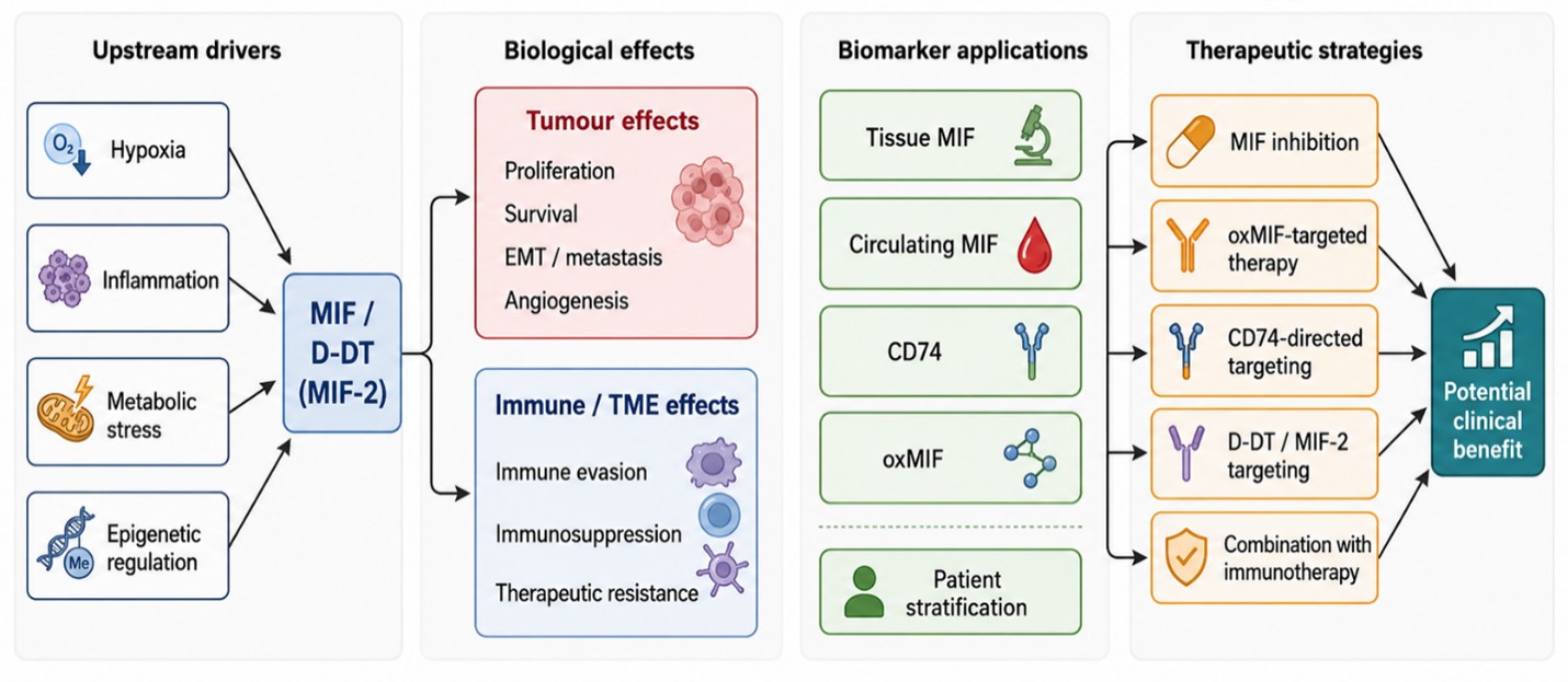
Fig. 3: Translational framework of MIF in oncology: from biology to biomarkers and therapy.
Biomarker Potential of MIF
Tissue expression and circulating MIF as diagnostic and prognostic biomarkers
Due to its detection in both tumor tissue and circulating compartments, MIF has become a potential biomarker candidate that can reflect biologically relevant aspects of tumor behavior. MIF serum levels have been studied in clinical trials involving genetic polymorphisms and breast cancer, which suggest the possible importance of circulating MIF in cancer biomarker studies (76). Likewise, serum MIF has been studied as a serological biomarker in oral squamous cell carcinoma, where serum-based measurement of MIF could give complementary information to the expression of MIF in local tissue (77). These findings, in combination, indicate that MIF might be useful for the stratification of tumors based on aggressiveness, tumor burden, or risk of progression. But the existing evidence is still exploratory, and the potential of MIF in clinical practice may rely on its combination with histopathological, molecular, and immune-related parameters.
Integrated biomarker panels, immune signatures, and barriers to adoption
While single-marker methods are still appealing, the biology of MIF signaling indicates that its most promising clinical applications might be in the context of larger molecular and immune-signature strategies. The tumor-immune context of the immunological tumor microenvironment may have a substantial impact on the biomarker potential of MIF-related pathways, as demonstrated by the proposal of MIF as a diagnostic and prognostic biomarker for TAMs M2 polarization in triple-negative breast cancer (78). Based on these observations, biomarkers related to MIF may be most informative when combined with other parameters in a multi-parameter model that characterizes the tumor/immune interaction more fully. This has been seen in other biomarker systems, including hypermethylation of NDRG4 in gastric cancer, where diagnostic and prognostic action requires integration with disease-specific molecular states (79). Thus, the main problems in using MIF-based biomarkers in clinical practice are biological heterogeneity, tumor specificity, standardization of assays, definition of cut-offs, and validating assays on independent patient cohorts. Thus, additional investigations will be required to validate MIF as a clinically useful biomarker in the field of oncology. The key therapeutic and biomarker significance of the MIF/CD74/DDT signaling axis in cancer is summarized in Table 3. Potential therapeutic strategies are summarized, such as the direct inhibition of MIF, the targeting of oxMIF, the targeting of CD74, D-DT/MIF-2 targeting, combinations with immunotherapy, and combinations with conventional anticancer therapy. Furthermore, it discusses the possible biomarker use of tissue expression of MIF, circulating MIF, oxMIF detection, CD74 expression, and combined immune-signature panels for prognosis, prediction of therapy response, and patient stratification.
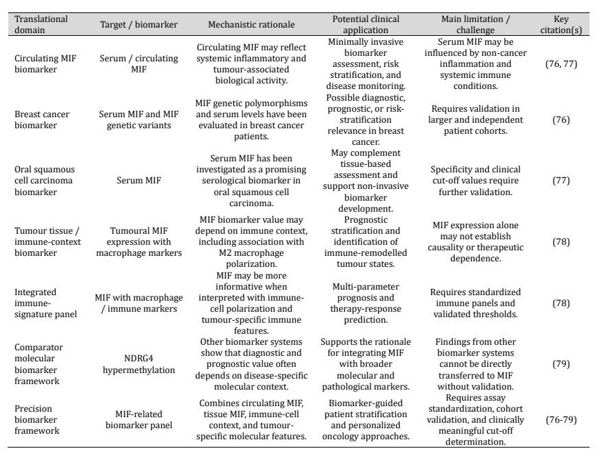
Table 3: Therapeutic and biomarker implications of MIF/CD74/DDT in cancer
Future Directions and Unresolved Challenge
Context dependence and cellular source of MIF signalling
One of the major unresolved questions in MIF biology is whether MIF signaling is uniformly pro-tumoral or whether it is very specific to microenvironmental context, cellular source, and stage of disease. This issue is particularly important because as MIF can be synthesized by tumor cells, stromal populations, and infiltrating immune cells, which may play distinct roles in tumor progression and remodeling of the tumor immune microenvironment. This is particularly complicated in metastatic disease or the central nervous system setting. The MIF-CD74 axis might have clinical relevance within the brain metastasis microenvironment, as pathway activity in these tumors seems to be tightly connected to cellular composition and tissue context (80). Similarly, studies in gliomas have shown that CD74 expression is largely restricted to microglia/macrophage populations and is associated with an M1-polarized immune milieu and longer patient survival, suggesting that MIF-receptor biology may vary according to cell type and immune context. It will thus be important to identify in future studies the source of the production of MIF, the respective cell populations responding to it, and the temporal change of the signaling activity in the premalignant, primary, and metastatic stages of the disease.Redundancy, causality, and limits of current evidence
Another major challenge in the field is that much of the current literature remains associative rather than definitively mechanistic. These studies often show a correlation with high MIF activity and aggressive disease behavior, immune suppression, and poor prognosis, but they do not necessarily provide insight into roles for MIF that are direct, co-regulatory, or downstream effects of tumor adaptation. The complexity of the receptor, redundancy of the signaling, and behavior of stress-responsive pathways further complicate this issue. MIF has been demonstrated to act as a noncognate ligand of the CXC chemokine receptors, meaning that MIF signaling is not restricted to the canonical CD74-mediated pathways and overlaps with chemokine-like receptor signaling systems (81). Moreover, experimental data in ER-positive breast cancer cells have demonstrated that chloroquine can promote ROS-mediated secretion of MIF and EMT, thereby revealing potential for treatment- or stress-related changes in MIF-related tumor-cell phenotypes (82). It will thus be important to perform more perturbation, longitudinal, and single-cell analyses in the future to delineate exactly how MIF acts mechanistically in various tumor microenvironments.
Precision translation through spatial biology, single-cell profiling, and biomarker-guided therapy
Translation of these advances will probably require better patient selection for modulation of MIF signaling as opposed to indiscriminate targeting of MIF signaling. The next step in research should be to identify tumor cell states that are particularly reliant on the pathways associated with MIF and to identify biomarkers that could be used to predict responsiveness to therapy. Single-cell RNA sequencing and spatial profiling will likely play a role in this process. In recent years, tumor heterogeneity in gastric carcinogenesis has attracted attention, and single-cell RNA sequencing has proven to be a valuable tool for investigating the existence of unique tumor states and how this correlates with tumor clinical parameters and could lead to more personalized therapeutic strategies (83, 84). Future research will thus need to combine spatial biology, single-cell profiling, biomarker-based patient stratification, and therapeutic studies to develop rational MIF-targeted treatment strategies. This could be important for separating the tumors where MIF is crucial for the tumor progression from those where MIF is part of the inflammatory, metabolic, or immune-microenvironmental adaptation process.
Conclusion
Macrophage migration inhibitory factor has emerged as a central regulator of inflammation-associated tumor progression and immune remodeling in cancer. The current evidence indicates that MIF is involved in various aspects of malignant progression, such as tumor-cell proliferation, survival, epithelial-mesenchymal transition, angiogenesis, microenvironmental remodeling, immune suppression, metabolic adaptation, and therapeutic resistance. Through its interactions with signaling partners such as CD74 and D-DT/MIF-2, the MIF signaling network appears to influence both tumor-intrinsic processes and the functional organization of the tumor microenvironment. Meanwhile, MIF's biological function is very situationally dependent. The effects of MIF signaling could be different depending on the type of tumor, receptor composition, cellular source, disease stage, and microenvironmental conditions. This complexity is likely one of the reasons for the high level of translational interest in MIF, and it is likely also responsible for the difficulties of targeting the pathway therapeutically. While several therapeutic strategies have been proposed through experimental studies, such as MIF inhibition, receptor-targeting strategies, and biomarker-guided combination therapy, many are still at the preclinical stage and need to be clinically validated. Future advances in the field will most likely rely on a better understanding of the spatial and single-cell distribution of MIF signaling, biomarker stratification, and incorporation of MIF-targeting strategies with current immunotherapeutic and anticancer treatments. The body of research indicates that MIF plays a significant role in immune system regulation and tumor microenvironment adaptability, and more mechanistic and clinically confirmed studies are needed to define the therapeutic usefulness of MIF in oncology.
Acknowledgements
The authors sincerely acknowledge Azerbaijan Medical University for providing the institutional support and academic environment that facilitated this work. The authors also extend their appreciation to departmental colleagues for their constructive discussions and intellectual input. Technical and administrative staff are thanked for their assistance during the preparation of the manuscript. Their collective support contributed significantly to the completion of this study.
Author Contributions
Conceptualization was undertaken by A.A. The original draft of the manuscript was prepared by A.A. and V.H. Review and editing were carried out by A.A. and M.N. Visualization was performed by A.A. and H.E. Supervision was provided by A.A. and G.K. Project administration was managed by A.A. and S.Y. Funding acquisition was secured by A.A. and G.S. All authors have read and approved the final version of the manuscript.
Disclosure Statement
The authors have no conflicts of interest to declare.
References
- Calandra T, Roger T. Macrophage migration inhibitory factor: a regulator of innate immunity. Nat Rev Immunol. 2003;3:791-800.https://doi.org/10.1038/nri1200
- Guda MR, Rashid MA, Asuthkar S, Jalasutram A, Caniglia JL, Tsung AJ, et al. Pleiotropic role of macrophage migration inhibitory factor in cancer. Am J Cancer Res. 2019;9:2760.
- Mora Barthelmess R, Stijlemans B, Van Ginderachter JA. Hallmarks of cancer affected by the MIF cytokine family. Cancers. 2023;15:395.https://doi.org/10.3390/cancers15020395
- Leng L, Metz CN, Fang Y, Xu J, Donnelly S, Baugh J, et al. MIF signal transduction is initiated by binding to CD74. J Exp Med. 2003;19711:1467-1476.https://doi.org/10.1084/jem.20030286
- Shi X, Leng L, Wang T, Wang W, Du X, Li J, et al. CD44 is the signaling component of the macrophage migration inhibitory factor-CD74 receptor complex. Immunity. 2006;254:595-606.https://doi.org/10.1016/j.immuni.2006.08.020
- Merk M, Zierow S, Leng L, Das R, Du X, Schulte W, et al. The D-dopachrome tautomerase gene product is a cytokine and functional homolog of macrophage migration inhibitory factor. Proc Natl Acad Sci U S A. 2011;10834: E577-E585.
- Valdez CN, Sánchez-Zuno GA, Bucala R, Tran TT. Macrophage migration inhibitory factor and D-dopachrome tautomerase: pathways to tumorigenesis and therapeutic opportunities. Int J Mol Sci. 2024;25:4849.https://doi.org/10.3390/ijms25094849
- Liu Z, Chen M, Zheng W, Yuan S, Zhao W. Insights into the prognostic value and immunological role of CD74 in pan-cancer. Discov Onc. 2024;15:222.https://doi.org/10.1007/s12672-024-01081-2
- Bozzi F, Mogavero A, Varinelli L, Belfiore A, Manenti G, Caccia C, et al. The MIF/CD74 axis is a target for novel therapies in colon carcinomatosis. J Exp Clin Cancer Res. 2017;361:16.https://doi.org/10.1186/s13046-016-0475-z
- Richard V, Kindt N, Saussez S. Macrophage migration inhibitory factor involvement in breast cancer. Int J Oncol. 2015;47:1627-1633.https://doi.org/10.3892/ijo.2015.3185
- Soumoy L, Kindt N, Ghanem G, Saussez S, Journe F. Role of macrophage migration inhibitory factor in melanoma. Cancers. 2019;11:529.https://doi.org/10.3390/cancers11040529
- Tanese K, Ogata D. The role of macrophage migration inhibitory factor family and CD74 in the pathogenesis of melanoma. Exp Dermatol. 2024;33:e15122.https://doi.org/10.1111/exd.15122
- Selo MA, Gobbo OL, Obaidi I, O'Connor C, Fayne D, Armstrong ME, et al. Bridging inflammation and oncology: the role and therapeutic potential of macrophage migration inhibitory factor in lung cancer. Int J Mol Sci. 2026;27:2829.https://doi.org/10.3390/ijms27062829
- Pu Y, Yang G, Zhou Y, Pan X, Guo T, Chai X. The macrophage migration inhibitory factor is a vital player in pan-cancer by functioning as an M0 macrophage biomarker. Int Immunopharmacol. 2024;134:112198.https://doi.org/10.1016/j.intimp.2024.112198
- Kindt N, Journe F, Laurent G, Saussez S. Involvement of macrophage migration inhibitory factor in cancer and novel therapeutic targets. Oncol Lett. 2016;12:2247-2253.https://doi.org/10.3892/ol.2016.4929
- O'Reilly C, Doroudian M, Mawhinney L, Donnelly SC. Targeting MIF in cancer: therapeutic strategies, current developments, and future opportunities. Med Res Rev. 2016;36:440-460.https://doi.org/10.1002/med.21385
- Wirtz TH, Saal A, Bergmann I, Fischer P, Heinrichs D, Brandt EF, et al. Macrophage migration inhibitory factor exerts pro-proliferative and anti-apoptotic effects via CD74 in murine hepatocellular carcinoma. Br J Pharmacol. 2021;178:4452-4467.https://doi.org/10.1111/bph.15622
- Rajasekaran D, Gröning S, Schmitz C, Zierow S, Drucker N, Bakou M, et al. Macrophage migration inhibitory factor-CXCR4 receptor interactions. J Biol Chem. 2016;291:15881-15895.https://doi.org/10.1074/jbc.M116.717751
- Zhang L, Woltering I, Holzner M, Brandhofer M, Schaefer CC, Bushati G, et al. CD74 is a functional MIF receptor on activated CD4+ T cells. Cell Mol Life Sci. 2024;81:296.https://doi.org/10.1007/s00018-024-05338-5
- Parkins A, Chen E, Rangel VM, Singh M, Xue L, Lisi GP, et al. Ligand-induced conformational changes enable intersubunit communications in D-dopachrome tautomerase. Biophys J. 2023;122:1268-1276.https://doi.org/10.1016/j.bpj.2023.02.019
- Tilstam PV, Pantouris G, Corman M, Andreoli M, Mahboubi K, Davis G, et al. A selective small-molecule inhibitor of macrophage migration inhibitory factor-2 inhibits MIF-2 biological activity. J Biol Chem. 2019;29449:18522-18531.https://doi.org/10.1074/jbc.RA119.009860
- Alban TJ, Grabowski MM, Otvos B, Bayik D, Wang W, Zalavadia A, et al. The MIF promoter SNP rs755622 is associated with immune activation in glioblastoma. JCI Insight. 2023;8:e160024.https://doi.org/10.1172/jci.insight.160024
- Chen T, Chen Q, Li F, Zeng M, Wang B, Huang S, et al. Targeted knockdown of macrophage migration inhibitory factor enhances UVB irradiation-induced apoptosis via increasing ROS generation in oral squamous cell carcinoma. Technol Cancer Res Treat. 2023;22:15330338231163436.https://doi.org/10.1177/15330338231163436
- Kindt N, Descamps G, Lechien JR, Remmelink M, Colet JM, Wattiez R, et al. Involvement of HPV infection in the release of macrophage migration inhibitory factor in head and neck squamous cell carcinoma. J Clin Med. 2019;8:75.https://doi.org/10.3390/jcm8010075
- Fan J, Ryu V, Qin Z, Dai L. Roles of macrophage migration inhibitory factor signaling pathway in oncovirus infection and virus-associated cancers. Viruses. 2025;17:1582.https://doi.org/10.3390/v17121582
- Osipyan A, Chen D, Dekker FJ. Epigenetic regulation in macrophage migration inhibitory factor-mediated signaling in cancer and inflammation. Drug Discov Today. 2021;26:1728-1734.https://doi.org/10.1016/j.drudis.2021.03.012
- Bandres E, Bitarte N, Arias F, Agorreta J, Fortes P, Agirre X, et al. microRNA-451 regulates macrophage migration inhibitory factor production and proliferation of gastrointestinal cancer cells. Clin Cancer Res. 2009;157:2281-2290.https://doi.org/10.1158/1078-0432.CCR-08-1818
- Tanese K, Hashimoto Y, Berkova Z, Wang Y, Samaniego F, Lee JE, et al. Cell surface CD74-MIF interactions drive melanoma survival in response to interferon-γ. J Invest Dermatol. 2015;13511:2775-2784.https://doi.org/10.1038/jid.2015.204
- Zhang JT, Zhang J, Wang SR, Yan LX, Qin J, Yin K, et al. Spatial downregulation of CD74 signatures may drive invasive component development in part-solid lung adenocarcinoma. iScience. 2023;26:107699.https://doi.org/10.1016/j.isci.2023.107699
- Li GQ, Xie J, Lei XY, Zhang L. Macrophage migration inhibitory factor regulates proliferation of gastric cancer cells via the PI3K/Akt pathway. World J Gastroenterol. 2009;15:5541.https://doi.org/10.3748/wjg.15.5541
- He XX, Chen K, Yang J, Li XY, Gan HY, Liu CY, et al. Macrophage migration inhibitory factor promotes colorectal cancer. Mol Med. 2009;15:1-10.https://doi.org/10.2119/molmed.2008.00107
- Klemke L, De Oliveira T, Witt D, Winkler N, Bohnenberger H, Bucala R, et al. Hsp90-stabilized MIF supports tumor progression via macrophage recruitment and angiogenesis in colorectal cancer. Cell Death Dis. 2021;12:155.https://doi.org/10.1038/s41419-021-03426-z
- Matsuura S, Shinmura K, Kamo T, Igarashi H, Maruyama K, Tajima M, et al. CD74-ROS1 fusion transcripts in resected non-small cell lung carcinoma. Oncol Rep. 2013;304:1675-1680.https://doi.org/10.3892/or.2013.2630
- Meyer-Siegler KL, Leifheit EC, Vera PL. Inhibition of macrophage migration inhibitory factor decreases proliferation and cytokine expression in bladder cancer cells. BMC Cancer. 2004;4:34.https://doi.org/10.1186/1471-2407-4-34
- Tan L, Ye X, Zhou Y, Yu M, Fu Z, Chen R, et al. Macrophage migration inhibitory factor is overexpressed in pancreatic cancer tissues and impairs the insulin secretion function of β-cells. J Transl Med. 2014;12:92.https://doi.org/10.1186/1479-5876-12-92
- Funamizu N, Hu C, Lacy C, Schetter A, Zhang G, He P, et al. Macrophage migration inhibitory factor induces epithelial-to-mesenchymal transition, enhances tumor aggressiveness, and predicts clinical outcome in resected pancreatic ductal adenocarcinoma. Int J Cancer. 2013;132:785-794.https://doi.org/10.1002/ijc.27736
- Fukuda Y, Bustos MA, Cho SN, Roszik J, Ryu S, Lopez VM, et al. Interplay between soluble CD74 and macrophage migration inhibitory factor drives tumor growth and influences patient survival in melanoma. Cell Death Dis. 2022;132:117.https://doi.org/10.1038/s41419-022-04552-y
- Huth S, Huth L, Heise R, Marquardt Y, Lopopolo L, Piecychna M, et al. Macrophage migration inhibitory factor and D-dopachrome tautomerase are significant promoters of UVB- but not chemically induced non-melanoma skin cancer. Sci Rep. 2023;13:11611.https://doi.org/10.1038/s41598-023-38748-9
- Liu W, Yang H, Zhi F, Feng Y, Luo H, Zhu Y, et al. Macrophage migration inhibitory factor may contribute to the occurrence of multiple primary lung adenocarcinomas. Clin Transl Med. 2023;13:e1368.https://doi.org/10.1002/ctm2.1368
- Liao Y, Wu C, Li Y, Wen J, Zhao D. MIF is a critical regulator of mononuclear phagocytic infiltration in hepatocellular carcinoma. iScience. 2023;26:107273.https://doi.org/10.1016/j.isci.2023.107273
- Yu M, Yu H, Wang H, Xu X, Sun Z, Chen W, et al. Tumor-associated macrophages activated in the tumor environment of hepatocellular carcinoma: characterization and treatment. Int J Oncol. 2024;65:100.https://doi.org/10.3892/ijo.2024.5688
- Liu W, Wang C, Liang L, Zhang C, Li Y, Xiao J, et al. Single-cell RNA sequencing analysis revealed the immunosuppressive remodeling of tumor-associated macrophages mediated by the MIF-CD74 axis in gastric cancer. Sci Rep. 2025;15:26883.https://doi.org/10.1038/s41598-025-10301-w
- Wu F, Feng Z, Wang X, Guo Y, Wu B, Bai S, et al. Sphingosine-1-phosphate stimulates colorectal cancer tumor microenvironment angiogenesis and induces macrophage polarization via macrophage migration inhibitory factor. Front Immunol. 2025;16:1564213.https://doi.org/10.3389/fimmu.2025.1564213
- Simpson KD, Templeton DJ, Cross JV. Macrophage migration inhibitory factor promotes tumor growth and metastasis by inducing myeloid-derived suppressor cells in the tumor microenvironment. J Immunol. 2012;18912:5533-5540.https://doi.org/10.4049/jimmunol.1201161
- Otvos B, Silver DJ, Mulkearns-Hubert EE, Alvarado AG, Turaga SM, Sorensen MD, et al. Cancer stem cell-secreted macrophage migration inhibitory factor stimulates myeloid-derived suppressor cell function and facilitates glioblastoma immune evasion. Stem Cells. 2016;348:2026-2039.https://doi.org/10.1002/stem.2393
- Ryan N, Lamenza F, Shrestha S, Upadhaya P, Springer A, Jordanides P, et al. Host-derived macrophage migration inhibitory factor expression attenuates anti-tumoral immune cell accumulation and promotes immunosuppression in the tumor microenvironment of head and neck squamous cell carcinoma. Biochim Biophys Acta Mol Basis Dis. 2024;1870:167345.https://doi.org/10.1016/j.bbadis.2024.167345
- Wang Z, Wang B, Feng Y, Ye J, Mao Z, Zhang T, et al. Targeting tumor-associated macrophage-derived CD74 improves the efficacy of neoadjuvant chemotherapy in combination with PD-1 blockade for cervical cancer. J Immunother Cancer. 2024;12:e009024.https://doi.org/10.1136/jitc-2024-009024
- Li D, Qiao B, Cheng Q, Chen L, Zhao F, Zuo L, et al. Macrophage migration inhibitory factor as a prognostic biomarker in multiple myeloma: clinical significance and in vitro effects. Carcinogenesis. 2025;46:bgaf033.https://doi.org/10.1093/carcin/bgaf033
- Pracha SH, Shrestha S, Ryan N, Upadhaya P, Lamenza FF, Jagadeesha S, et al. Targeting macrophage migration inhibitory factor to inhibit T-cell immunosuppression in the tumor microenvironment and improve cancer outcomes in head and neck squamous cell carcinoma. Oral Oncol. 2025;160:107126.https://doi.org/10.1016/j.oraloncology.2024.107126
- Tran TT, Sánchez-Zuno GA, Osmani L, Caulfield J, Valdez CN, Piecychna M, et al. Improving immunotherapy responses by dual inhibition of macrophage migration inhibitory factor and PD-1. JCI Insight. 2025;10:e191539.https://doi.org/10.1172/jci.insight.191539
- Bonnin E, Rodrigo Riestra M, Marziali F, Mena Osuna R, Denizeau J, Maurin M, et al. CD74 supports the accumulation and function of regulatory T cells in tumors. Nat Commun. 2024;15:3749.https://doi.org/10.1038/s41467-024-47981-3
- Liu X, Song J, Yuan M, Zuo F, Li H, Tang L, et al. Single-cell transcriptional dissection illuminates an evolution of immunosuppressive microenvironment during pancreatic ductal adenocarcinoma metastasis. Signal Transduct Target Ther. 2025;10:182.https://doi.org/10.1038/s41392-025-02265-0
- Yang S, Tang W, Azizian A, Gaedcke J, Ohara Y, Cawley H, et al. MIF/NR3C2 axis regulates glucose metabolism reprogramming in pancreatic cancer through MAPK-ERK and AP-1 pathways. Carcinogenesis. 2024;45:582-594.https://doi.org/10.1093/carcin/bgae025
- Wang X, Li D, Zhu B, Hua Z. Single-cell transcriptome analysis identifies a novel tumor-associated macrophage subtype predicting better prognosis in pancreatic ductal adenocarcinoma. Front Cell Dev Biol. 2024;12:1466767.https://doi.org/10.3389/fcell.2024.1466767
- Ringel AE, Drijvers JM, Baker GJ, Catozzi A, García-Cañaveras JC, Gassaway BM, et al. Obesity shapes metabolism in the tumor microenvironment to suppress anti-tumor immunity. Cell. 2020;1837:1848-1866.https://doi.org/10.1016/j.cell.2020.11.009
- Yao Y, Luo L, Xiang G, Xiong J, Ke N, Tan C, et al. The expression of m6A regulators correlated with the immune microenvironment, which plays an important role in the prognosis of pancreatic ductal adenocarcinoma. Gland Surg. 2022;11:147-165.https://doi.org/10.21037/gs-21-859
- Kaymak I, Williams KS, Cantor JR, Jones RG. Immunometabolic interplay in the tumor microenvironment. Cancer Cell. 2021;391:28-37.https://doi.org/10.1016/j.ccell.2020.09.004
- Jarmula J, Lee J, Lauko A, Rajappa P, Grabowski MM, Dhawan A, et al. Macrophage migration inhibitory factor as a therapeutic target in neuro-oncology: a review. Neurooncol Adv. 2024;6:vdae142.https://doi.org/10.1093/noajnl/vdae142
- Winner M, Meier J, Zierow S, Rendon BE, Crichlow GV, Riggs R, et al. A novel macrophage migration inhibitory factor suicide substrate inhibits the motility and growth of lung cancer cells. Cancer Res. 2008;6818:7253-7257.https://doi.org/10.1158/0008-5472.CAN-07-6227
- Shun CT. Expression of macrophage migration inhibitory factor is associated with enhanced angiogenesis and advanced stage in gastric carcinomas. World J Gastroenterol. 2005;11:3767.https://doi.org/10.3748/wjg.v11.i24.3767
- Huang Z, Wu C, Liu X, Lu S, You L, Guo F, et al. Single-cell and bulk RNA sequencing reveal malignant epithelial cell heterogeneity and prognosis signatures in gastric carcinoma. Cells. 2022;11:2550.https://doi.org/10.3390/cells11162550
- Pan M, Chen P, Zhang Q, Yang Y, Hu W, Hu G, et al. Comprehensive profiling of cell subsets of gastric cancer and liver metastasis based on single-cell RNA-sequencing analysis. Transl Cancer Res. 2024;13:330-347.https://doi.org/10.21037/tcr-23-1532
- Kurahara H, Shinchi H, Mataki Y, Maemura K, Noma H, Kubo F, Sakoda M, Ueno S, Natsugoe S, Takao S. Significance of M2-polarized tumor-associated macrophage in pancreatic cancer. Journal of Surgical Research. 2011 May 15;1672:e211-9. https://doi.org/10.1016/j.jss.2009.05.026https://doi.org/10.1016/j.jss.2009.05.026
- Imaoka M, Tanese K, Masugi Y, Hayashi M, Sakamoto M. Macrophage migration inhibitory factor-CD74 interaction regulates the expression of programmed cell death ligand 1 in melanoma cells. Cancer Sci. 2019;110:2273-2283.https://doi.org/10.1111/cas.14038
- Thiele M, Donnelly SC, Mitchell RA. OxMIF: a druggable isoform of macrophage migration inhibitory factor in cancer and inflammatory diseases. J Immunother Cancer. 2022;10:e005475.https://doi.org/10.1136/jitc-2022-005475
- Puchol Tarazona AA, Schinagl A, Mirkina I, Rossmueller G, Kerschbaumer RJ, Bachmann F, et al. Pretargeted radioimmunotherapy with the novel anti-oxMIF/HSG bispecific antibody ON105 results in significant tumor regression in murine models of cancer. Mol Cancer Ther. 2024;23:1219-1229.https://doi.org/10.1158/1535-7163.MCT-24-0083
- Schneider KL, Claus L, Bucala R, Schulz-Heddergott R. Targeting macrophage migration inhibitory factor as a potential therapeutic strategy in colorectal cancer. Oncogenesis. 2025;14:30.https://doi.org/10.1038/s41389-025-00572-3
- Wang L, Shen G, Xie G, Li Z, Ma X, Li M, et al. Targeting mast cell activation and MIF-mediated remodelling enhances chemotherapy response in pancreatic cancer. Adv Sci. 2025;12:e09930.https://doi.org/10.1002/advs.202509930
- Lee SH, Kwon HJ, Park S, Kim CI, Ryu H, Kim SS, et al. Macrophage migration inhibitory factor inhibitor 4-IPP downregulates stemness phenotype and mesenchymal trans-differentiation after irradiation in glioblastoma multiforme. PLoS One. 2021;169:e0257375.https://doi.org/10.1371/journal.pone.0257375
- Jeong H, Lee SY, Seo H, Kim BJ. Recombinant Mycobacterium smegmatis delivering a fusion protein of human macrophage migration inhibitory factor and IL-7 exerts an anticancer effect by inducing an immune response against MIF in a tumor-bearing mouse model. J Immunother Cancer. 2021;9:e003180.https://doi.org/10.1136/jitc-2021-003180
- Liu L, Wang J, Wang Y, Chen L, Peng L, Bin Y, et al. Blocking the MIF-CD74 axis augments radiotherapy efficacy for brain metastasis in NSCLC via synergistically promoting microglia M1 polarization. J Exp Clin Cancer Res. 2024;43:128.https://doi.org/10.1186/s13046-024-03024-9
- Mangano K, Mazzon E, Basile MS, Di Marco R, Bramanti P, Mammana S, et al. Pathogenic role for macrophage migration inhibitory factor in glioblastoma and its targeting with specific inhibitors as a novel tailored therapeutic approach. Oncotarget. 2018;9:17951-17970.https://doi.org/10.18632/oncotarget.24885
- Sajko S, Skeens E, Schinagl A, Ferhat M, Mirkina I, Mayer J, et al. Redox-dependent plasticity of oxMIF facilitates its interaction with CD74 and therapeutic antibodies. Redox Biol. 2024;75:103264.https://doi.org/10.1016/j.redox.2024.103264
- Pantouris G, Bucala R, Lolis EJ. Structural plasticity in the C-terminal region of macrophage migration inhibitory factor-2 is associated with an induced fit mechanism for a selective inhibitor. Biochemistry. 2018 May 30;5726:3599-605. https://doi.org/10.1021/acs.biochem.8b00344https://doi.org/10.1021/acs.biochem.8b00344
- Takahashi A, Iwata T, Yamashita K. Knockdown of D-dopachrome tautomerase inhibits cell proliferation in human HepG2 cell line. Anticancer Res. 2021;41:4077-4082.https://doi.org/10.21873/anticanres.15209
- Seliem MA, Mohamadin AM, El-Sayed MIK, Ismail Y, El-Husseiny AA. The clinical signature of genetic variants and serum levels of macrophage migration inhibitory factor in Egyptian breast cancer patients. Breast Cancer Res Treat. 2024;208:57-66.https://doi.org/10.1007/s10549-024-07393-9
- Zepeda-Nuño JS, Gutiérrez-Cortés E, Hernández-Bello J, Ángeles-Sánchez J, de la Cruz-Mosso U, Cruz Á, et al. Macrophage migration inhibitory factor: a promising oncogenic serological biomarker for oral squamous cell carcinoma. Int J Immunopathol Pharmacol. 2021;35:20587384211038417.https://doi.org/10.1177/20587384211038417
- Chen M, Liu H, Hong B, Xiao Y, Qian Y. MIF as a potential diagnostic and prognostic biomarker for triple-negative breast cancer that correlates with the polarization of M2 macrophages. FASEB J. 2024;38:e23696.https://doi.org/10.1096/fj.202400578R
- Chen X, Yang Y, Liu J, Li B, Xu Y, Li C, et al. NDRG4 hypermethylation is a potential biomarker for diagnosis and prognosis of gastric cancer in the Chinese population. Oncotarget. 2017;8:8105-8119.https://doi.org/10.18632/oncotarget.14099
- Cai H, Lu X, Fan W, Fang C, Mu F, Bai H, et al. Single-cell combined with bulk transcriptomics reveals cross-cancer common regulatory mechanisms in the brain metastasis microenvironment and the clinical significance of the MIF-CD74 axis. Genes Dis. 2026;102178.https://doi.org/10.1016/j.gendis.2026.102178
- Zeiner PS, Preusse C, Blank A, Zachskorn C, Baumgarten P, Caspary L, et al. MIF receptor CD74 is restricted to microglia/macrophages, associated with an M1-polarized immune milieu and prolonged patient survival in gliomas. Brain Pathol. 2015;25:491-504.https://doi.org/10.1111/bpa.12194
- Bernhagen J, Krohn R, Lue H, Gregory JL, Zernecke A, Koenen RR, et al. MIF is a noncognate ligand of CXC chemokine receptors in inflammatory and atherogenic cell recruitment. Nat Med. 2007;135:587-596.https://doi.org/10.1038/nm1567
- Rojas-Sánchez G, García-Miranda A, Montes-Alvarado JB, Cotzomi-Ortega I, Sarmiento-Salinas FL, Jiménez-Ignacio EE, et al. Chloroquine induces ROS-mediated macrophage migration inhibitory factor secretion and epithelial-to-mesenchymal transition in ER-positive breast cancer cell lines. J Mammary Gland Biol Neoplasia. 2021;26:341-355.https://doi.org/10.1007/s10911-021-09503-5
- Xu J, Yu B, Wang F, Yang J. Single-cell RNA sequencing to map tumor heterogeneity in gastric carcinogenesis, paving roads to individualized therapy. Cancer Immunol Immunother. 2024;73:233.https://doi.org/10.1007/s00262-024-03820-4