Original Article – DOI: 10.33594/000000879
CPB (60): 408 - 416
Accepted: 16.07.2026 - Published: 31.07.2026
Genotype–Phenotype Correlation of Β-Globin Mutations and Hematological Parameters in Iraqi Β-Thalassemia Patients
Keywords
Abstract
Background/Aims: β-Thalassemia is a common hemoglobin disorder distributed worldwide and caused by mutations in the HBB gene that reduce or abolish β-globin chain synthesis, resulting in chronic hemolytic anemia, ineffective erythropoiesis, and variable clinical severity. Although the molecular spectrum of β-thalassemia has been extensively investigated, data correlating β-globin mutations with hematological characteristics in Iraqi patients remain limited. This study aimed to evaluate genotype–phenotype correlations by integrating molecular mutation analysis with hematological and biochemical profiling in an Iraqi β-thalassemia cohort. Methods: A cross-sectional study was conducted between September and November 2025 at the Hematology Department, University of Baghdad. Fifty patients with β-thalassemia were initially recruited, of whom 22 fulfilled the inclusion criteria and consented to participate (4 males and 18 females; age range, 18–68 years). Hematological parameters, HbA₂ and HbF levels, serum iron and ferritin concentrations, and β-globin gene mutations were analyzed. Mutation screening was performed using amplification refractory mutation system polymerase chain reaction (ARMS-PCR). Statistical analyses were conducted to evaluate genotype–phenotype associations. Results: Patients exhibited a predominantly microcytic, hypochromic hematological profile, with a mean hemoglobin concentration of 11.1 ± 1.4 g/dL, a mean MCV of 63.5 ± 0.78 fL, a mean MCH of 20.2 ± 0.25 pg, and elevated RDW values (17.9 ± 0.23%), consistent with anisocytosis. HbA₂ levels ranged from 3.5% to 7.2% (mean, 5.4 ± 0.25%) as determined by HPLC, whereas HbF levels ranged from 0.3% to 5.6% (mean, 1.4 ± 0.17%). HPLC provided more reliable HbA₂ quantification than alkaline electrophoresis. Serum iron and ferritin levels showed considerable interindividual variability. β-Globin mutations were identified in 68.2% of patients, with IVS-I-6 being the most frequent mutation (45.5%), followed by CD39. No significant associations were observed between genotype and sex, profession, ethnicity, or place of residence. In contrast, age-group distribution differed significantly. Conclusion: This study demonstrated considerable phenotypic heterogeneity among Iraqi patients with β-thalassemia and identified IVS-I-6 as the predominant β-globin mutation in this cohort. The absence of significant genotype–phenotype associations for most clinical variables suggests that additional genetic and environmental modifiers may contribute to disease expression. Combined molecular and hematological evaluation may improve the diagnostic characterization and classification of patients with β-thalassemia.
Introduction
Thalassemia represents a group of inherited hemoglobin disorders caused by the reduced or absent synthesis of one or more globin chains, leading to chronic anemia and a spectrum of clinical complications [1, 2]. It is considered a major global public health concern, especially in the Mediterranean region and the Middle East, where it has a relatively high prevalence. Blood transfusion remains the cornerstone of management for patients with severe disease; however, it is associated with progressive iron overload and an increased risk of transfusion-transmitted infections [3, 4]. In Iraq, β-thalassemia is considered one of the most common inherited blood disorders [5, 6]. β-Thalassemia is caused by mutations in the HBB gene that reduce or alter β-globin chain synthesis, resulting in an imbalance between α- and β-globin chain production, ineffective erythropoiesis, chronic hemolysis, and variable clinical severity [7]. Advances in molecular diagnostics, especially allele-specific amplification techniques such as amplification refractory mutation system polymerase chain reaction (ARMS-PCR), have substantially improved the detection and characterization of HBB mutations and facilitated genotype-based diagnosis [8]. Clinical severity varies considerably among patients and is influenced not only by the underlying mutation but also by genetic and environmental modifiers. The prognosis of the disease is closely related to the severity of anemia and the age at clinical presentation [9]. Regular transfusion therapy, iron chelation, and hematopoietic stem cell transplantation have improved survival and quality of life; however, considerable phenotypic heterogeneity remains among affected individuals [10]. Despite the high prevalence of β-thalassemia in Iraq, comprehensive studies integrating molecular mutation analysis with hematological characterization remain limited. Therefore, the present study aimed to investigate the relationship between HBB gene mutations and hematological parameters in Iraqi patients with β-thalassemia, providing region-specific molecular and clinical data that may contribute to improved diagnostic characterization and patient classification.
Materials and Methods
Study Design and Patient Population
This cross-sectional study was conducted between September and November 2025 at the Hematology Department, University of Baghdad, Iraq. Fifty patients diagnosed with β-thalassemia were initially selected. After applying the inclusion and exclusion criteria, 22 patients, including 4 males and 18 females, aged between 18 and 68 years, were selected for the final analysis. Patients were distributed into three age groups (≤18 years, 18–40 years, and >40 years). The inclusion criteria included adults diagnosed with β-thalassemia according to hematological findings and hemoglobin electrophoresis showing HbA₂ levels greater than 3.5%, with or without elevated HbF levels. Patients who had received a blood transfusion within the previous three months were excluded to avoid the influence of transfused blood on hematological and electrophoretic parameters. A healthy control group (n = 18), matched for age and sex, was also included. Demographic and clinical information, including age, sex, medical history, medication, and other relevant clinical parameters, were obtained from patients' medical records. Before blood collection, all participants were informed about the purpose of the study and provided written informed consent.
Blood Collection and DNA Extraction
Peripheral venous blood (5 mL) was collected into EDTA-containing vacuum tubes. Samples were kept refrigerated until laboratory processing. Peripheral blood leukocytes were used for genomic DNA extraction using a commercial DNA extraction kit according to the manufacturer's instructions. DNA concentration and purity were assessed spectrophotometrically before molecular analysis.
β-Globin Mutation Analysis
β-globin gene mutation screening was performed using amplification refractory mutation system polymerase chain reaction (ARMS-PCR) with allele-specific primers targeting the five most common Mediterranean and Middle Eastern β-globin mutations detected in this population: IVS-I-6 (T→C), IVS-I-1 (G→A), IVS-I-5 (G→C), IVS-I-110 (G→A), and CD39 (C→T). Each 25-µL PCR reaction contained genomic DNA, mutation-specific and control primers, dNTPs, MgCl₂, reaction buffer, and Taq DNA polymerase. PCR amplification consisted of an initial denaturation at 95°C for 5 min, followed by 30 cycles of denaturation at 95°C for 30 s, primer annealing at 58–62°C for 30 s, and extension at 72°C for 30 s, with a final extension at 72°C for 7 min. Amplified products were separated by agarose gel electrophoresis and visualized under UV illumination following ethidium bromide staining. Mutation-specific bands were evaluated using both a normal control and a no-template control. Among the identified compound heterozygous mutations, one patient carried IVS-I-6 (T→C)/IVS-I-5 (G→A), two patients carried IVS-I-6 (T→C)/IVS-I-5 (G→C), one patient carried IVS-I-6 (T→C)/IVS-I-110 (G→A), and one patient carried IVS-I-1 (G→A)/IVS-II-849 (A→G). Homozygous IVS-I-6 mutations were observed in patients with intermediate β-thalassemia, whereas patients with minor β-thalassemia exhibited greater mutation heterogeneity.
Osmotic Resistance Test
The osmotic resistance test was carried out as a screening method for β-thalassemia, especially in heterozygous individuals, since microcytic erythrocytes are more resistant to osmotic hemolysis. Although approximately 97% of heterozygous β-thalassemia patients showed a positive result, the test is not specific because positive findings may also be observed in patients with hemoglobinopathies (AS, AC, SS, SC, and CC), α-thalassemia, and iron deficiency anemia. A 10% NaCl stock solution at pH 7.4 was prepared, and a 0.36% NaCl working solution was subsequently obtained for testing.
Hemoglobin Electrophoresis
Under alkaline conditions, hemoglobin electrophoresis was carried out for the separation and semi-quantification of normal and abnormal hemoglobin fractions based on their electrophoretic mobility. Differences in migration were observed due to alterations in the electrical charge of hemoglobin molecules caused by amino acid substitutions within the globin chains. An automated SPIFE/REP electrophoresis system (Helena Laboratories, Beaumont, TX, USA) was used for sample application, electrophoretic separation, staining, destaining, gel drying, densitometric analysis, image acquisition, and result interpretation.
High-Performance Liquid Chromatography (HPLC)
HbA₂ quantification was additionally performed by high-performance liquid chromatography (HPLC). In this technique, the mobile phase continuously transports the sample through a chromatographic column (stationary phase). Differences in retention time allow the separation and quantitative measurement of individual hemoglobin fractions following calibration with appropriate standards.
Statistical Analysis
Statistical analyses were performed using the Statistical Analysis System (SAS, 2018). The software was used to compare study groups and evaluate associations between demographic, molecular, and hematological variables. Statistical significance was determined using the methods described for each analysis, and a P value of <0.05 was considered statistically significant.
Results
Severe forms of β-thalassemia are managed with three main approaches: frequent blood transfusions, iron chelation to remove excess iron, and splenectomy, the need for which increases with transfusion frequency. Only 22 of the fifty patients with β-thalassemia agreed to participate in the study. Although 50 patients were initially recruited, only 22 were included due to refusal, incomplete data, or a recent blood transfusion within the previous three months. Of these 22 patients, 4 were men and 18 were women. The ethnicity of the study population was predominantly mixed, with a small minority identifying as White (2 of 22 patients, 9.1%). The professions included domestic work, and five patients were students; the remainder held various occupations, including farming, craftwork, nursing assistance, sewing, clerical work, driving, production operation, and teaching. Most patients affected by the disease were from Baghdad. Table 1 shows the reference values for the biochemical and hematological parameters studied. The hematological parameters showed wide variability among the studied samples. Erythrocyte counts ranged from 3.92 to 7.39 million/mm³ (mean: 5.5 ± 0.11), while hemoglobin levels ranged from 8.5 to 15.3 g/dL (mean: 11.1 ± 1.4) and hematocrit values ranged from 27.5% to 47.3% (mean: 34.9 ± 4.04), indicating the presence of mild to moderate anemia in a proportion of cases. Red blood cell indices demonstrated a predominantly microcytic and hypochromic pattern, with reduced MCV (54.4–71.8 fL; mean: 63.5 ± 0.78) and MCH (17.3–22.1 pg; mean: 20.2 ± 0.25), while MCHC remained relatively stable (29.8–33.6 g/dL; mean: 31.8 ± 0.12). Elevated RDW-CV (14.7–20.6%; mean: 17.9 ± 0.23) and RDW-SD (31.3–44.6 fL; mean: 38.7 ± 0.83) reflected anisocytosis. Hemoglobin fraction analysis showed increased HbA₂ levels (3.5–7.2%; mean: 5.4 ± 0.25 by HPLC, and 3.5–6.8%; mean: 5.2 ± 0.17 by electrophoresis), with elevated HbF levels (0.3–5.6%; mean: 1.4 ± 0.17). Biochemical analysis showed a wide range of serum iron (27.0–217.0 µg/dL; mean: 98.9 ± 5.86) and ferritin levels (5–1804 ng/mL; mean: 244.7 ± 324.92), indicating marked interindividual variability in iron status among participants, with no significant differences in serum iron or ferritin levels between patient subgroups. No statistically significant associations were found between disease phenotype (minor versus intermediate β-thalassemia) and sex (P = 0.625), profession (P = 0.727), or ethnicity (P = 1.000). Patients from the capital showed a numerically higher proportion of intermediate β-thalassemia compared with those from interior regions, but this difference did not reach statistical significance (P = 0.078), as shown in Table 2. Table 3 presents the age distribution of the β-thalassemia cohort, with a mean age of 44.8 years (range: 18–68 years), and most patients (68.2%) falling within the >40-year age group. The age-group distribution differed significantly (P = 0.036). The comparison between HbA₂ levels measured by the two different methods (HPLC and alkaline electrophoresis) is shown in Table 4. By HPLC, HbA₂ ranged from 3.5% to 7.2% (mean: 5.4 ± 0.25%), whereas by alkaline electrophoresis it ranged from 3.5% to 6.8% (mean: 5.2 ± 0.17%). Table 5 presents the demographic, hematological, and molecular characteristics of patients with β-thalassemia minor (denoted "TM" in the table). Most individuals exhibited mild anemia with moderately elevated HbA₂ and low HbF levels. At the molecular level, IVS-I-6 and CD39 were the predominant mutations identified, while a subset of patients had no identifiable mutation. Table 6 describes the demographic and hematological features, in addition to the molecular characteristics, of patients with intermediate β-thalassemia. These patients showed more severe anemia, with lower hemoglobin levels and higher HbF and HbA₂ percentages compared with those with the minor form. Homozygous IVS-I-6 mutations were frequent. The mutation distribution of β-thalassemia according to phenotype showed that the IVS-I-6 mutation was present in 10 (45.5%) of the 68.2% of patients with a characterized mutation. Five patients had a clinical diagnosis of intermediate β-thalassemia, while the other five had a clinical diagnosis of minor β-thalassemia. Table 7 shows that mutation type was not significantly associated with the measured clinical traits.
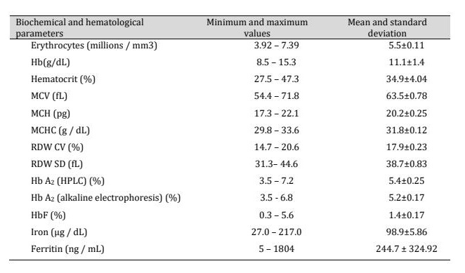
Table 1: Reference values, mean, standard deviation, and minimum and maximum values for biochemical and hematological parameters
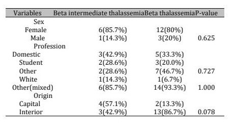
Table 2: Association between demographic characteristics (sex, profession, ethnicity, and place of residence) and β-thalassemia phenotype (minor vs. intermediate) in the study cohort
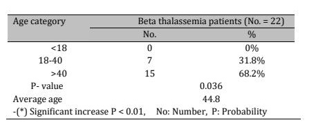
Table 3: the distribution of age groups between patients with beta thalassemia
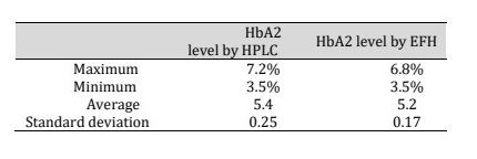
Table 4: The comparison between HbA2 level in β-thalassemia patients using the HPLC and electrophoresis in alkaline medium method
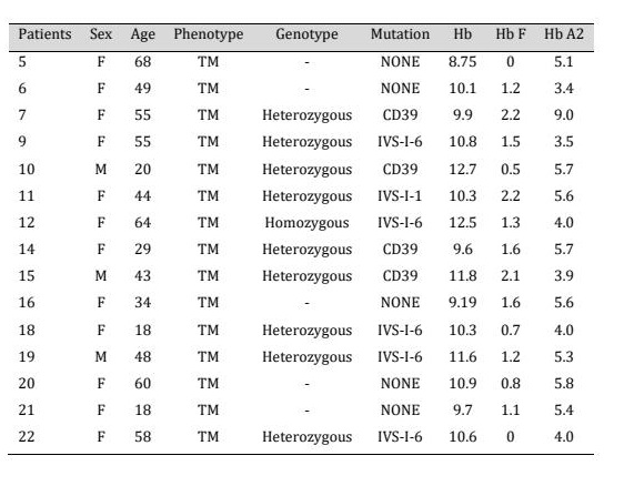
Table 5: Demographic and hematological features of individuals with beta-thalassemia minor. TM = thalassemia minor
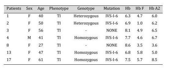
Table 6: Demographic and hematological features of individuals with intermediate beta thalassemia. TI = thalassemia intermediate
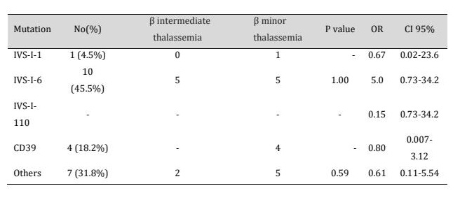
Table 7: Individuals with beta thalassemia are distributed according to the kind of mutation and their phenotype
Discussion
The findings of the present study highlight the substantial clinical and molecular phenotypic heterogeneity of β-thalassemia within the examined Iraqi cohort, reflecting the multifactorial nature of disease expression [11]. The significant, non-uniform distribution of patients across age groups observed in this cohort (Table 3) likely reflects differences in survival, timing of diagnosis, or referral patterns between the minor and intermediate disease forms rather than a true epidemiological pattern. The hematological profile, characterized by microcytosis, hypochromia, elevated RDW, and anisocytosis, was fully consistent with the underlying pathophysiology of ineffective erythropoiesis secondary to defective β-globin chain production [12]. HbA₂ levels above 3.5%, measured by both alkaline electrophoresis and HPLC, represented an essential diagnostic marker for β-thalassemia [13]. This finding is consistent with previous studies indicating the superior sensitivity of HPLC over alkaline electrophoresis for HbA₂ quantification in the screening and diagnosis of heterozygous β-thalassemia. The HPLC data obtained in the present study support its preferential use in diagnostic settings, especially where coexisting iron deficiency or anemia could lead to confounding interpretation. Serum iron and ferritin levels, although heterogeneous across the cohort, did not indicate that the observed hematological abnormalities were primarily attributable to iron deficiency rather than β-globin chain imbalance. At the molecular level, IVS-I-6 was the predominant mutation, followed by CD39 and other variants, consistent with previously reported Iraqi mutation spectra [1]. The examination of the genotype–phenotype relationship in this cohort showed that patients homozygous for IVS-I-6 were concentrated in the intermediate β-thalassemia group and tended to present with lower hemoglobin levels and higher HbF levels, as highlighted in Tables 5 and 6, consistent with the known association between homozygous IVS-I-6 and a more severe clinical phenotype. In contrast, heterozygous genotypes, including both CD39 and IVS-I-6 heterozygosity, were distributed across both the minor and intermediate phenotypes, with substantial overlap in hematological indices. In addition, several patients with intermediate disease carried no identifiable mutation within the screened panel. This trend indicates that the primary β-globin genotype alone does not fully predict disease severity in this population, as shown in Table 7, and that the broad phenotypic overlap between genotype subgroups is more consistent with a continuum of disease severity than with discrete genotype-defined categories. However, the absence of a statistically significant genotype–phenotype relationship underscores that β-thalassemia severity cannot be explained solely by the primary β-globin mutation. The current findings support a role for secondary genetic modifiers, such as HbF persistence and co-inherited globin variants, consistent with recent reports describing the distribution of genetic modifiers affecting hemoglobin expression [14]. Additionally, environmental and therapeutic factors, such as transfusion exposure, adherence to iron chelation therapy, and possibly oxidative stress pathways, may also contribute to disease severity. The observed overlap between the minor and intermediate β-thalassemia phenotypes supports the concept of β-thalassemia as a clinical continuum rather than a rigid categorical disorder. The findings of the present study are consistent with previous studies by Mohammed and colleagues, who reported reduced MCV and MCH values in patients with β-thalassemia. Furthermore, elevated HbA₂ levels (>3.5%) were consistent with the established diagnostic criteria reported by Ling [13,15]. The predominance of the IVS-I-6 mutation was also consistent with previous studies conducted in Middle Eastern populations [7].
Conclusion
It is important to highlight that the current results represent a region-specific contribution by integrating hematological, biochemical, and molecular findings within a single analytical framework, thereby improving the understanding of the diversity of β-thalassemia in Iraqi patients. However, genotype–phenotype interpretation was limited by several factors, including incomplete baseline clinical information, such as transfusion burden, longitudinal iron overload progression, splenectomy status, and chelation regimens. This study of β-thalassemia in an Iraqi cohort demonstrated considerable phenotypic diversity despite the presence of identifiable β-globin mutations, indicating that genotypic variation alone may be insufficient to predict clinical expression. The combined application of detailed hematological indices, accurate HbA₂ quantification by HPLC, and molecular characterization can strengthen diagnostic accuracy and improve patient classification.
Acknowledgements
All authors contributed significantly to the conception and design of the study, data collection, analysis, and manuscript preparation. No specific funding was received for this study. No artificial intelligence tools were used in the design of the study. The study protocol was reviewed and approved by the Research Ethics Committee of the University of Baghdad, Institute of genetic engineering and biotechnology for post graduate, Baghdad, Iraq under the reference number (CSEC/1023/0086 in September 29, 2025.). Written informed consent was obtained from all participants before enrolment. The datasets generated and/or analyzed during the current study are available from the corresponding author upon reasonable request.
Disclosure Statement
The authors have nothing to disclose.
References
- Mohammed SJ, Al-Lami F, Hussein MQ: Electrocardiographic changes among beta-thalassemic major patients in Ibn Al-Baladi Thalassemia Center-Baghdad. Al-Kindy Coll Med J 2013;9(1):36-41.
- Sadiq IZ, Abubakar FS, Usman HS, Abdullahi AD, Ibrahim B, Kastayal BS, Ibrahim M, Hassan HA: Thalassemia: pathophysiology, diagnosis, and advances in treatment. Thalass Rep 2024;14(4):81-102. https://doi.org/10.3390/thalassrep14040010
- Ali ZH: Prevalence and risk factors for hepatitis C virus in beta-thalassemic patients attending blood diseases center in Ibn Al-Baladi Hospital, Baghdad. Al-Kindy Coll Med J 2018;14(1):42-49. https://doi.org/10.47723/kcmj.v14i1.17
- Almuktar AA, Al Obaidy LHA, Ali AM: The impact of VDR-FokI polymorphism in Iraqi patients with prostate cancer and prostate benign hyperplasia. Baghdad Sci J 2025;22(3):Article 14.
- Hassan R, Al-Mendalawi M: Growth failure in Iraqi children with β-thalassemia major. Iraqi J Med Sci 2019;20:41-48.
- Majid M, Mutar M: Thalassemia prevention program in Iraq: will it be cost-effective and applicable. Baghdad Med J Stud 2020;1 DOI: 10.48046/bmjos.v1i1.2. https://doi.org/10.48046/bmjos.v1i1.2
- Zhang B, Yang Y, Zhu Y, Zhang S, Huang H, Sun Q: The discovery of hemoglobin body: a start to more. Precis Nutr 2024;3(2):00071. https://doi.org/10.1097/PN9.0000000000000071
- Ahmed HM, Georgy DB, Meabed MH, Abd El Kareem RM, Botrous OE: The association between plasma microRNA-451 expression levels and chronic kidney disease in children with β-thalassemia major. Iran J Kidney Dis 2022;16(3):188-94.
- Keith J, Christakopoulos GE, Fernandez AG, Yao Y, Zhang J, Mayberry K, Telange R, Sweileh RBA, Dudley M, Westbrook C, Sheppard H, Weiss MJ, Lechauve C: Loss of miR-144/451 alleviates β-thalassemia by stimulating ULK1-mediated autophagy of free α-globin. Blood 2023;142(10):918-32. https://doi.org/10.1182/blood.2022017265
- Amin S, Jalal S, Ali K, Mohammed A, Rasool L, Osman T: Beta-thalassemia intermedia: a single thalassemia center experience from Northeastern Iraq. Biomed Res Int 2020;2020:1-11. https://doi.org/10.1155/2020/2807120
- Abbas RA, Hassan RH, Taghlubee IM, Mohammed SI, Mohammed HH, Hasan HH, et al: Prevalence and molecular characterization of β-thalassemia in Kirkuk Province, Northern Iraq. Hemoglobin 2024;48(5):308-13. https://doi.org/10.1080/03630269.2024.2418507
- Khaleel FM, Muhi SA, Selman AD: Assessment of pro-hepcidin and its relation with iron profile in hemodialysis male patients. Baghdad Sci J 2016;13(1):Article 11. https://doi.org/10.21123/bsj.2016.13.1.0074
- Ling L, Wang F, Li Y, He S, Wu F, Yang L, Xu L, Wang T, Zhou S, Yang F, Wei Z, Yang L, Yang Z, Fang X, Zhou Y, Xue J, Yin X, Wei H, Yu D: Depletion of miR-144/451 alleviates anemia in β-thalassemic mice. Blood Adv 2024;8(10):2565-70. https://doi.org/10.1182/bloodadvances.2022008757
- Diamantidis MD, Ikonomou G, Argyrakouli I, Pantelidou D, Delicou S: Genetic modifiers of hemoglobin expression from a clinical perspective in hemoglobinopathy patients with beta thalassemia and sickle cell disease. Int J Mol Sci 2024;25(22):11886.https://doi.org/10.3390/ijms252211886
- Aljanabi, Noor H. and Abed, Reema M. (2024) "Investigation the role of Human Leukocyte Antigen-G in Iraqi patients with papillary thyroid carcinoma," Baghdad Science Journal: Vol. 21: Iss. 6, Article 18. https://doi.org/10.21123/bsj.2023.8695